Showing posts with label Photos. Show all posts

Tuesday, August 28, 2018

Cracking the Sugar Code

Cracking the sugar code: Why the 'glycome' is the next big thing in health and medicine
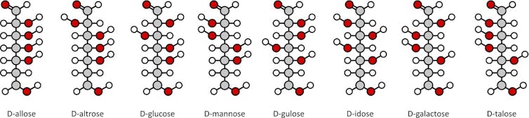
File 20180724 194149 1a8cg12.jpg?ixlib=rb 1.1
By molekuul_be/shutterstock.com
Emanual Maverakis, University of California, Davis; Carlito Lebrilla, University of California, Davis, and Jenny Wang, Yeshiva University

When you think of sugar, you probably think of the sweet, white, crystalline table sugar that you use to make cookies or sweeten your coffee. But did you know that within our body, simple sugar molecules can be connected together to create powerful structures that have recently been found to be linked to health problems, including cancer, aging and autoimmune diseases.

These long sugar chains that cover each of our cells are called glycans, and according to the National Academy of Sciences, creating a map of their location and structure will usher us into a new era of modern medicine. This is because the human glycome – the entire collection of sugars within our body – houses yet-to-be-discovered glycans with the potential to aid physicians in diagnosing and treating their patients.

Thanks to the worldwide attention garnered by the 2003 completion of the Human Genome Project, most people have heard about DNA, genomics and even proteomics – the study of proteins. But the study of glycans, also known as glycomics, is about 20 years behind that of other fields. One reason for this lag is that scientists have not developed the tools to rapidly identify glycan structures and their attachment sites on people’s cells. The “sugar coat” has been somewhat of a mystery.

Until now, that is.

While most laboratories focus on cellular or molecular research, our lab is dedicated to developing technology to rapidly characterize glycan structures and their attachment sites. Our ultimate goal is to catalog the hundreds of thousands of sugars and their locations on various cell types, and then to use this information to tailor medical therapies to each individual.

Why do we care about glycans?

In the future, it is likely that analysis of an individual’s glycans will be used to predict our risk for developing diseases like rheumatoid arthritis, cancer or even food allergies. This is because glycome alterations can be specifically tied to particular disease states. Also, biological processes like aging are linked to inflammation in our glycome. It remains to be tested if reversing these changes can help prevent disease, or even slow aging – an intriguing possibility.

Along with DNA, proteins, and fats, glycans are one of the four major macromolecules essential for life. Of these four, glycans are the final arbiters of how our cells behave.

DNA orchestrates what we look like, our capacity to think and behave, and even determines the diseases to which we are most susceptible. Within our DNA are short segments, genes, which often contain instructions for how to synthesize proteins. Proteins in turn are the “workhorses” of the cell, carrying out many of the functions necessary for life.

However, how a protein behaves often depends on what glycans are attached to it. In other words, these sugar molecules can greatly influence how our proteins do their work, and even how our cells will respond to stimuli. For example, if you change a few glycans on the outside of a cell, it might trigger that cell to migrate to a different location in our body.

The main job of glycans is to modify the proteins and fats that sit on the surface of our cells. Together, they create a thick sugar coat around the cell. If we consider the surface of the cell to be soil, then glycans would be the wonderfully diverse plant-life and foliage that sprout up and bring color and identity to the cell. In fact, if you were able to see a cell with your naked eye, it would look very fuzzy. Picture a peach with 10 times more fuzz.
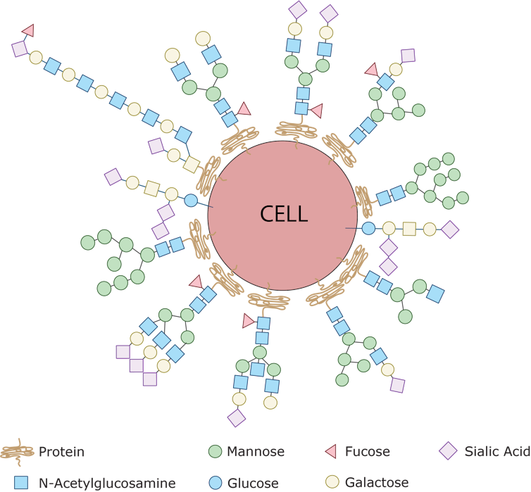
Every single cell in the human body is covered with a collection of glycans which are assembled using various simple sugars like glucose, mannose, galactose, sialic acid, glucosamine and frucose as building blocks. By sensing the type of sugar coat present, our immune cells can identify other cells as friend or foe. This is because bacteria have sugars on their surfaces that are never seen on human cells – the pathogen’s sugars are sensed by the immune system and that identifies the bacteria as ‘foreign.’ Emanual Maverakis, CC BY-SA

Glycans label our own cells and identify them as ‘self’

The fuzz around a cell is its glycan coat. Being on the outside of our cells, glycans are the first point of contact for most cellular interactions and thus influence how our cells communicate with one another. You can also think of the glycans as a unique cellular “barcode.” Thus, a kidney cell’s fuzz will look different from an immune cell’s fuzz. But there are also similarities. In fact, the immune cells that survey our body searching for pathogens know not to attack our own “self” cells because of common features in the glycan “barcode” which are shared by all cells of our body.

In contrast, bacteria and parasites like malaria have different “sugar coats” that are not seen on human cells. When bacterial sugars are tagged as “foreign,” a person’s immune system targets the bacterium for destruction. However, some harmful bacterial pathogens like group B streptococcus, which commonly cause severe infections in babies, can avoid immune detection by impersonating human cells by carrying similar glycans as a disguise – like the wolf dressed in sheepskin.

Unfortunately some pathogens are also able to use our glycans to help them cause disease. Deadly viruses like HIV and Ebola have evolved to grab hold of specific glycans which they then “lock” onto as they infect our human cells. Therapies that either block these viruses from interacting with our glycans, or that attack virus-specific glycans may be a new avenue to treating these infections.
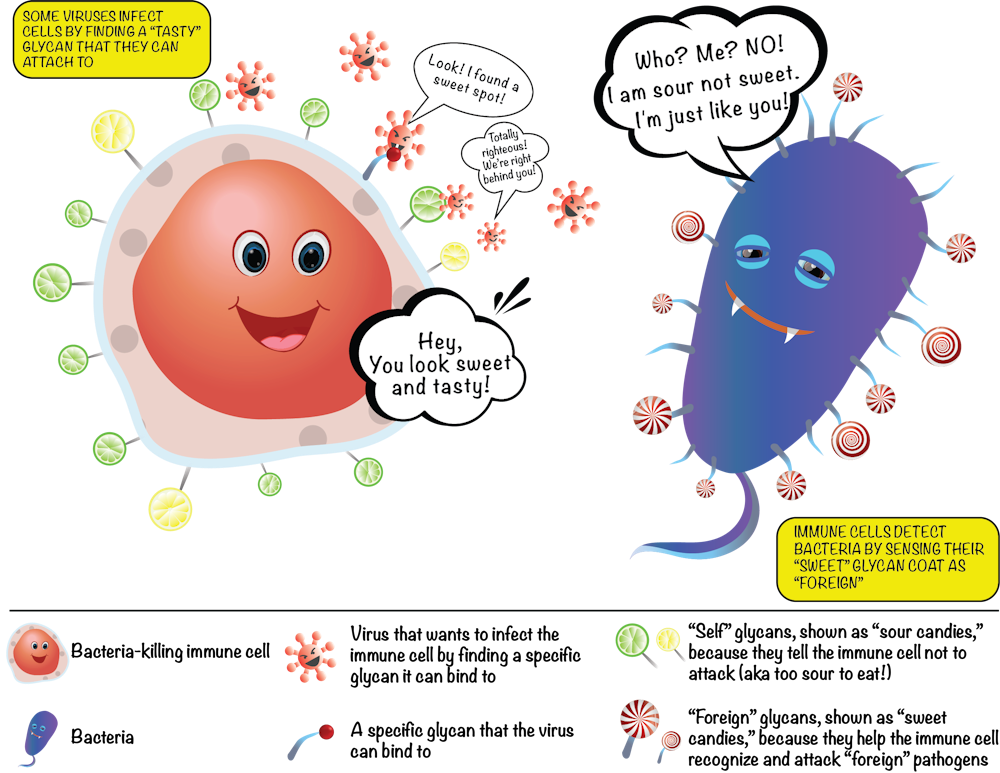
The sugars on our cells and on bacterial cells label them as friend or foe. Emanual Maverakis, CC BY-SA

New research has also shown that glycans play a huge role in the development of autoimmune diseases like rheumatoid arthritis and autoimmune pancreatitis. This is not surprising since glycans directly influence the function of immune cells.

Normally, our immune cells act as our body’s “defense system,” and identify and destroy foreign invaders like harmful bacteria or viruses. But when the body mistakenly labels our own cells as the enemy and launches an internal attack on itself, autoimmunity is born. Interestingly, in such instances, it is the glycans present on the misbehaving self-attacking antibodies that will dictate the strength of the attack on the body. This abnormal immune response can even be directed against glycans. For example, the immune system can mistake “self” glycans as if they were “foreign” molecules. Our research team recently published an article that introduced the glycan theory of autoimmunity, which explains some of these relationships.

Glycans in our food can trigger immune responses

There have been many studies linking consumption of red meat with diseases like atherosclerosis and diabetes, but they have not been able to show why or how this occurs until recently. One intriguing study suggests that the culprit was a sugar with the unwieldy name, nonhuman sialic N-glycolylneuraminic acid, or Neu5Gc for short. Neu5Gc is found in all mammals except humans, because the early humans that could make Neu5Gc died from an ancient malarial parasite.

However, although we now lack the ability to produce Neu5Gc, our bodies still have the ability to incorporate it into the glycans on our cells if we obtain it by eating red meat. Once it becomes part of our cells’ glycan coat, our cells then have a “foreign” substance – Neu5Gc – surrounding them. This can trigger inflammation throughout the body because our immune system recognize Neu5Gc as “foreign” and attacks it. The chronic inflammation caused by these internal attacks can lead to heart attack, stroke and even cancer.

Our bodies synthesize tens of thousands of unique glycans, often with branching structures formed from simple sugar building blocks. Proteins or fats can also be modified by dozens of unique glycans. These countless combinations make mapping glycans a difficult task because we need a practical and efficient way to analyze hundreds of thousands of glycan patterns.

Our research team has now developed methods to rapidly and robustly monitor the human glycome. By capitalizing on engineering advancements and improvements in sample processing, our technique can monitor thousands of glycans at once, which allows us to characterize the glycans in cells from healthy controls and patients with a variety of different diseases. Our goal is to use this data to develop predictive models to help clinicians diagnose and treat all human diseases. We believe that a new wave of medical advancements will arrive as we unlock the “sugar code.”

Jenny Wang was the co-lead author of this article.The Conversation

Emanual Maverakis, Associate Professor- Departments of Medical Microbiology & Immunology and Dermatology | Member- Foods For Health Institute | Member- Comprehensive Cancer Center | Director- Autoimmunity | Director- Immune Monitoring Core, University of California, Davis; Carlito Lebrilla, Distinguished Professor of Chemistry, University of California, Davis, and Jenny Wang, Clinical Research Fellow, University of California, Davis | Medical Student, Albert Einstein College of Medicine, Yeshiva University

This article was originally published on The Conversation. Read the original article.

Wednesday, February 21, 2018

Pyoderma Gangrenosum- Diagnosis


  • Pyoderma gangrenosum (PG), which can be associated with rheumatoid arthritis and inflammatory bowel disease, is a difficult disease to diagnose and treat.  Traditionally PG has been considered a "diagnosis of exclusion" but the disease has several characteristic features.  These include pathergy-> PG ulcers often appear at a sites of minor trauma.  Another characteristic of the disease is the rapid evolution of the lesions-> they first appear as papules or pustules and then they rapidly ulcerate. On clinical exam cribriform or "wrinkled paper" scars at sites of healed ulcers can also aid in the diagnosis of PG.  The ulcers themselves have a zone of peripheral erythema, undermined borders and are very tender.  Unfortunately PG is often misdiagnoses or over diagnosed leading to mismanagement of one of the most devastating dermatologic diseases.  A group of international experts recently came together to develop diagnostic criteria for this disease.  It is the hope that these criteria will help aid physicians in diagnosing PG and clinical researchers conduct well-designed clinical trials. This research was recently published in JAMA Dermatology.  Please see the following link.
  • Diagnostic Criteria of Ulcerative Pyoderma Gangrenosum: A Delphi Consensus of International ExpertsJAMA Dermatol. Published online February 14, 2018. doi:10.1001/jamadermatol.2017.5980


Thursday, February 8, 2018

Gamma Delta T cells protect against Staph aureus


Gamma Delta T cells protect against Staph aureus

Need to characterize an immune response? My research team specializes in developing novel bioinformatic methods to analyze RNA-Seq datasets for immune genes and in performing dedicated T cell receptor gene sequencing. Take a look at our recent article in the Journal of Clinical Investigation, which demonstrates how gamma delta T cells provide long-term protection against Staph aureus. This project was based out of Lloyd Miller's laboratory at Johns Hopkins University.  Dr. Miller is an expert in Staph aureus and wanted to the study anti-Staph immune responses.  For Dr. Miller our research team used our in-house developed bioinformatics pipeline (TCRminer) to correlate the expression of T cell receptor genes with other immune genes of interest.  We also performed dedicated TCR sequencing to answer fundamental questions about how the immune system responds to Staph.  Please see the link to the article and please contact us if this type of analysis can be of use in your research. We are especially interested in immune monitoring in the setting of immunotherapy and autoimmunity. Collaborative studies are most welcome.

Friday, August 25, 2017


National Psoriasis Foundation's Facebook Live event to promote their Treat-to-Target philosophy on psoriasis care featured an interview with Dr. Maverakis and his patient Howard Chang.


Click on image to watch the video.


 Here is a link to the video

Sunday, July 2, 2017

UC Davis Health hosts migrant student workshop


Each year our research team helps out the Migrant Student Leadership Institute, a program hosted by the College Assistance Migrant Program (CAMP) by brining migrant students to UC Davis.  This year we brought 145 Californian migrant students to UC Davis for a day of talks, tours, and research activities.  Medical students, graduate students and professors participated in the event.

For more on this year's event read the UC Davis press release (click here).


Jason Kao's white coat ceremony.  All of these clinical researchers are off to bigger and better things.  Michelle and Chelsea are starting dermatology residency at UC Davis.  Jason Koo is now in PA school and Elizabeth Wang (<-far left) is applying to dermatology residency and she is off on her away rotations. 

Wednesday, March 8, 2017

Elaine Fuchs


Elaine Fuchs, PhD was nice enough to visit UC Davis to give a lecture and spend a little time with our research team.  Dr. Fuchs is known for numerous discoveries, including her work characterizing the different keratin genes and the genetic defects that they mutations in them cause.  Her lab has been on the forefront of a variety of discoveries for the past several decades.  She is a recipient of the National Medal of Science and a Howard Hughes Investigator.

Pellegra and Psoriasis, an interesting combination.


Our star medical student Elizabeth Wang presented her poster at the AAD this week. It was on a case presentation of a patient with pellegra and psoriasis. This is an interesting disease combination because both disease produce psoriasis-(like) changes on pathology but psoriasis gets better with phototherapy while pellegra is exacerbated by UV radiation. This was one of the clues that led to the second diagnosis of pellegra in this patient.  He ended up responding well to niacin. (Dr. Maverakis is a member of the world renown UC Davis Foods for Health Institute). Elizabeth was mentored by Dr. Cindy Chambers on this project.

Sunday, February 19, 2017

Dr. Youn Kim was nice enough to visit our clinical trials group and sit down with us during her recent trip to UC Davis.  Dr. Kim is an esteemed clinician-educator and researcher.  She is Professor of Dermatology and the Director of Stanford's Multidisciplinary Cutaneous Lymphoma Clinic/Program.

Saturday, May 14, 2016

CBS News and Dr. Maverakis


Click here to watch the CBS news report- my 2 seconds of fame

CBS, Sacramento Bee, and other news outlets exaggerate the strength of the protests against Katehi by showing old footage and by trying to encourage students to continue protesting.  The truth is that only a tiny fraction of the more than 33,000 UC Davis students are protesting the chancellor.  None showed up at the UC Regents meeting despite being encouraged by the news media to do so.

The majority of UC Davis faculty support Chancellor Katehi and are upset at Janet Napolitano for her political attack against the chancellor.  A biased investigation will prove nothing.  Recently the NAACP has also voiced their concern about how the allegations against Chancellor Katehi are being handled.

CBS shows only a few seconds of the faculty speeches to the UC Regents but conducts long interviews with a few students protestors who did not even show up the UC Regents's meeting.


Monday, March 7, 2016

Florida Study Section

Dr. Maverakis returns from a CSR study section in Florida.  No alligator sitings but he did run into some interesting wildlife.  


Sunday, March 6, 2016

Dr. Maverakis Public Lecture

Dr. Maverakis lecturing to the public on strategies to maintain good skin health (approximately 520 people in attendance).  Click the photo to see more photos.


Sunday, January 24, 2016

UCSF Grandrounds

Dr. Maverakis gives Grand Rounds at UCSF.  


His basic science presentation covered the role of glycans in the immune system. A second clinical talk was on intralesional therapy for in transit melanoma metastases.  After his presentations, Dr. Maverakis had the opportunity to meet with talented young researchers and faculty.  The short drive down to UCSF allowed him to meet new dermatologists and see old friends.

Thursday, January 14, 2016

Taiwan Trip

Dr. Maverakis giving a keynote address at Taipei Medical University.  CLICK THE PHOTO TO SEE MORE PHOTOS!


The trip allowed him to reconnect with old friends and meet new collaborators.  

Wednesday, December 16, 2015

Elani Linos


Dermatologist and Public Health Leader Dr. Eleni Linos Comes to Visit the Maverakis Lab.

We had a fun two days with Eleni Linos, MD, DrPH.  Dr. Linos is a dually trained in public health and dermatology.  She is a leader in the field of skin cancer research and prevention. During her stay she gave an amazing grand rounds presentation.

Altered Glycans and Autoimmunity


 Altered Glycans and Autoimmunity


LINK TO ARTICLE

Howard Chang, a columnist for everyday Health, was nice enough to write an article about our research on how glycans (oligosaccharides) are linked to autoimmunity.  Please take a look at his very nice piece.

Thursday, September 17, 2015

Thuy Phung, MD

Dermatopathologist, physician-scientist and global health leader Thuy Phung comes to visit the Maverakis Lab

We had a fun and enlightening 3 days with her.   Dr. Phung is one of the founders of an award-winning dermatology clinic in Vietnam that specializes in treating children with vascular malformations. She is also faculty at the Baylor College of Medicine.  Her laboratory has made seminal discoveries on how abnormal blood vessels grow.  She came to visit the Maverakis Lab and also give Grand Rounds at UC Davis.


Saturday, August 29, 2015

Dr. Maverakis on CBS News

Dr. Maverakis meets with CBS Channel 13 to talk about Drinkable sunscreens. 


Carotenoids and other components of drinkable sunscreens can provide a minimal amount of sun protection but should not be considered a replacement for your topical sunscreen.